
Circular Staplers
* Esophagectomy,Gastrectomy,Small intesting and
Colon Excision Operation,Rectum low antrior resection
Linear Cutter Staplers
* General Surgery, Chest Surgery,Gynecology
and the parenchyma Excision,transection,reforger and anastomose of pediatric Operation.
Skin staplers offer a variety of surgical, closure application
Veterinary surgeons have largely embraced surgical stapling, especially during the past 10 years. The reasons are apparent to those veterinarians familiar with surgical stapling equipment: reduction in surgery/anesthesia time for the patient, simplicity of application and consistency in results. Some doctors have been slow to embrace surgical stapling, primarily because of the potential cost concerns associated with investing in the equipment and supplies, a lack of training experience and a limited surgical caseload in a given practice.
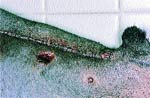 Photo 1: Skin staplers provide a convenient and safe way to close surgical wounds. |
From my clinical experience, there are two occasions where skin staples may not be ideally suited for wound closure: closing wounds under moderate tension, and use on thin skin. Conventional tension suture patterns are better able to maintain wound closure in the face of moderate incisional tension, especially in areas where regional motion can enhance this distracting force. Thin skin does not always provide sufficient dermal thickness for the proper seating or embedding of skin staples. As a result, the skin staples have a tendency to tip or rotate, making their subsequent removal problematic. Surprisingly, skin glue applied over the staple line, can reduce or eliminate this tendency, while providing an additional "footprint" of security.
In more recent years, skin staplers have been effectively used to secure skin grafts to the cutaneous tissue bordering the recipient site. The skin graft is applied over the recipient site, with its borders slightly overlapping the wound edge. Skin staples are applied while adjusting the graft tension to assure proper graft to bed contact. Skin staples also are used to secure dressings over a grafted area to prevent shifting of the dressing beneath the overlying bandage layers.
Skin staples are stainless steel in composition. As a result, they can be effectively used as internal radiographic markers when applied to the subcutaneous tissues and underlying muscle fascia after tumor resection. These metallic markers can assist the radiation oncologist in determining the margins of the surgical field and the appropriate region to irradiate postoperatively.
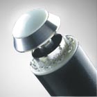
Vascular staplers Vascular clips are an invaluable method to rapidly ligate small bleeding vessels during surgical procedures. Improvements in the design of the disposable applicators have made their clinical use much more "user friendly." The Kendall/USSC Surgiclip units, for example, currently offered have an automatic staple loader to facilitate rapid placement of flat vascular clips.
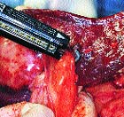 Photo 2: A ligate-divide-staple is ideal to use for splenectomies. |
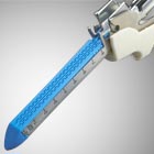
Linear stapling devices Linear staplers apply two to four rows of fine titanium staples in a staggered configuration. Thoracoabdominal (TA) staplers apply two rows of staples; the exception is the TA-30 V-3 cartridge that seals vascular pedicles with a triple row of staples (for added security). Gastrointestinal anastomosis (GIA) staplers apply four rows of staples. The most popular GIA cartridges contain a cutting blade that divides between the second and third rows of staples during application. As a result, the GIA and TA staplers have somewhat different uses.
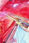 Photo 4: Gastrointestinal anastomosis staplers can be effective in resecting large areas of compromised gastric tissue. |
In combination with the GIA stapler, functional end-to-end anastomosis of the intestinal tract can be performed in dogs and the larger feline patients. Similarly, they also can be employed for cholecystoduodenostomy, cholecystojejunostomy and the Bilroth II procedures.
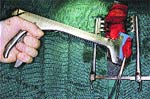 Photo 3: Thoracoabdominal staplers apply two rows of staples and are best used with partial or complete lung lobectomies. |
Other considerations Smaller versions of the tissue staplers are available for use in endoscopic surgery. Although this area of small animal surgery is limited in scope at this time, it is an important area in human surgery. These modified instruments can be used for minimally invasive surgery, or applied in combination with conventional approaches to the thorax and abdomen, especially in smaller veterinary patients.
The newer designs in stapling equipment continue to facilitate their application in surgery. Over time, veterinarians will continue to use staplers for conventional applications, while determining new uses for this innovative line of instruments.
Dr. Pavletic is the head of the Department of Surgery and director of surgical services at Boston's Angell Memorial Animal Hospital. He is a 1974 graduate of the University of Illinois and a 1981 diplomate of the American College of Veterinary Surgeons. He is the author of the 1999 Atlas of Small Animal Reconstructive Surgery, Second Edition, published by W.B. Saunders.
More informatin Come From www.medicalstapler.com
